
|
|
|
"Use of a Squatting Movement as a Clinical Marker of Function After Total Knee Arthroplasty"1/10/2013  With skyrocketing health care costs, insurance agencies are constantly looking for ways to reduce reimbursement. This includes decreasing payments to organizations that have poor outcomes. As most PTs are looking to stay in business, it is important that we have valid and reliable outcome measures in order to receive appropriate reimbursement. This study looked at the effectiveness of analyzing the symmetry of a squat as a clinical marker of function after Total Knee Arthroplasty. As many functional activities involve a squat-like maneuver, the squat may prove to be a valid measure for functional ability. The authors compared body weight % on each side in standing, 30 degree squats, and 60 degree squats. For anyone who has seen a knee replacement surgery, it's easy to understand why individuals place the majority of their weight on the uninvolved limb following surgery. The compensations caused by the asymmetries place the uninvolved limb and joints around the involved knee at risk for additional injuries due to their increased load. The study found that the 60 degree squat asymmetries was correlated with individuals who had moderate difficulty with functional activities, while standing asymmetries were not. Symmetry was found to be improved upon further rehabilitation. The squat may be a useful outcome measure when treating patients following total knee replacements. Upon further research, this may be a common tool used in the clinic for assessing patient progression.
1 Comment
 In the previous post, we discussed a clinical prediction rule for patients presenting with neck pain who would benefit from thoracic manipulation.
We would like to clarify that the CPR only provides the ability to a priori identify individuals who would likely have early success after being treated with thoracic spine manipulation. A priori is defined as reasoning that proceeds from theoretical deduction rather than experience or observation. Cleland et al stated in their original article that further studies needed to be performed in order to validate this CPR. A follow-up study found that this CPR did not prove valid. However, Cleland et al did point out that while the CPR lost validity, patients with mechanical neck pain who were treated with thoracic spine manipulation had significantly greater improvements in short and long term disability (based off the Neck Disability Index). Additionally, these individuals had less pain at 1-week follow up compared with individuals who only received exercise. In conclusion, the CPR for patients presenting with neck pain who would benefit from thoracic manipulation was not validated. As clinicians, still be cognizant that thoracic manipulation can improve your patient's neck pain and disability status. While the 6 predictive variables of the CPR cannot be used in isolation, the importance of these factors should still be documented to help guide your differential diagnosis. A quick thank you to Steve who brought this information to our attention! Neck Disability Index criteria for scoring can be found HERE.  This is an excellent article that reviews exercise physiology, the body's response to exercise, and long-term adaptation. It does a quick summary of basic information like muscle types, metabolic response (aerobic vs anaerobic), and the activities that activate each level of metabolism. One of the initial responses to exercise is the cardiovascular response. At first, stroke volume increases alone, but at 40-60% of the VO2max, cardiac output can only increase through increased heart rate. Tissues can also improve their oxygen content by increasing oxygen extraction from the blood (arterial-venous difference). Of course, blood pressure will be found to increase as well (systolic). Additionally, hydration is a well-known component of exercise and can have significant impact on exercise performance. Dr. Noakes, on the other hand, presents the opinion of hydration levels being overly criticized as the culprit in Lore of Running. His research has found hypoglycemia and hyponatremia to be the cause of decreased athletic performance far more frequently. Regular endurance exercise has been shown to have several effects on the body. Muscles have increased mitochondria size and capillary formation. This increases blood flow to the muscle and the cells' ability to extract/utilize oxygen. Plasma volume has also been shown to increase, thus, improving the viscosity of the blood, making it easier for it to flow through the body. The increased work load results in left ventricular hypertrophy, which leads to stronger contractions. For much more, check out the article yourself!
Reference: Rivera-Brown AM, Frontera WR. Principles of Exercise Physiology: Responses to Acute Exercise and Long-term Adaptations to Training (2012). PM&R: The Journal of Injury, Function, and Rehabilitation 4(11), 797-804. An essential clinical requirement is having valid and reliable Range of Motion measurements. This objective data is important not only to differentiate patients, but also for reimbursement purposes. Here is a quick review on landmarks, fulcrum placement, normal end feel, and normal Range of Motion using goniometric measurement. While not all clinics use these same anatomical landmarks, familiarizing yourself with the general concepts is important! *Much of the information was taken from the Norkin and White Textbook. 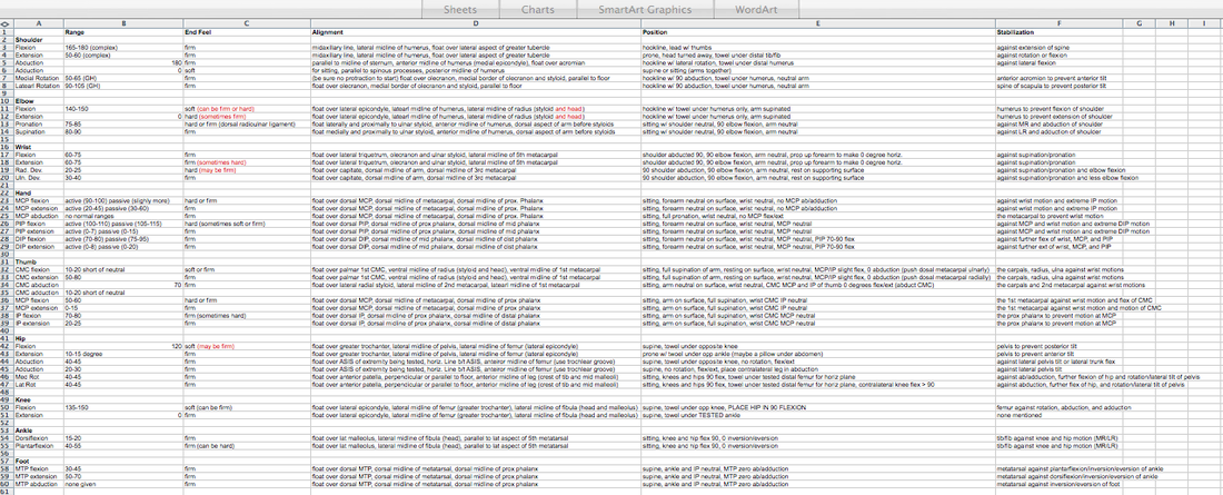
As students, we are fortunate to have access to Pubmed, Iliad, and a variety of other professional databases. The newest information in the field is spoon fed to us by our teachers. If we have questions, we can easily use the resources we are paying for through our education. As we graduate, many of these resources will be taken away. How will you continue stay on top of all the new evidence? This short youtube video gives a quick look at how to stay current with research. Additionally, Mike Reinold discusses several other strategies to stay up to date in the profession of physical therapy.
How to Read Research Articles: http://www.mikereinold.com/2011/04/how-to-read-research-articles.html 3 Steps to Integrate Evidence Based Medicine Into What You Do: It Is Not That Hard: http://www.mikereinold.com/2011/01/3-steps-to-integrate-evidence-based-medicine-into-what-you-do-it-is-not-that-hard.html  Background Information:
Biceps Tendinitis encompasses a spectrum of disorders including primary biceps tendinitis, patients without primary tendinitis, and biceps tendinosis. Primary Biceps Tendinitis (5%) is inflammation of the biceps tendon in the intertubercular groove caused by mechanical stresses and overuse. Some clinicians identify biceps tendinitis simply by individuals who exhibit anterior shoulder pain that is exacerbated by shoulder and elbow flexion (Mitra et al, 2011). Mechanical stresses can include entrapment, instability, and spontaneous rupture of the long head of the biceps tendon. These patients often suffer from secondary impingement because of scapular instability, anterior capsule laxity, or posterior joint capsule tightness. Patients suffering from secondary impingement are often your younger athletic population between 18-35 years old. Primary tendinitis is much less common than those without primary tendonitis (95%). The patients without primary tendonitis have biceps tendon pathology secondary to a rotator cuff injury or labral tear. Interestingly, biceps tendinopathy is rarely found alone. It usually occurs secondary to or a cause of another pathology (Zhang et al, 2011). 85% of patients with long head of the biceps tendon partial tears have associated rotator cuff pathologies (Gazillo 2011). Additionally the umbrella term biceps tendinitis includes biceps tendinosis, which is caused by primary impingement of the shoulder and overuse. This usually involves your older population >35 years of age. Due to their age and primary impingement, these patients are also at risk for rotator cuff pathology. CLICK HERE TO CONTINUE READING Some practitioners may think they'll never encounter a patient with lymphedema based on the setting. With the fragility of the lymphatic system, a patient can develop lymphedema with surgery, treatment, trauma, and more, relatively easily. With the impedance it can place on the patient's rehabilitation, it is imperative we are aware of the best treatment methods regarding lymphedema so that we can acceleration the patient's healing process. This study reviews the evidence behind Complete Decongestive Therapy (CDT). CDT is performed daily until tissue normalization occurs. It consists of: - ~60 minutes daily of Manual Lymph Drainage (MLD) - Multi-layer, short-stretch compression bandaging with foam or layers of fabric padding of the affected limbs - Exercises to enhance lymphatic pumping - Skin care of affected areas - Compression garments for maintenance following reductions 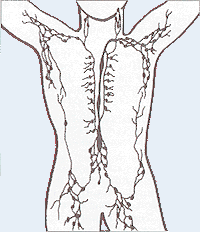 The lymphatic system is made up of a collection of lymph vessels and nodes that transport a protein rich fluid known as lymph. After undergoing trauma, the lymphatic system has the potential to become overwhelmed. Either there is too much lymph on the body or the lymphatic system can no longer support the load. This leads to the pathology known as lymphedema. The increase in fluid impairs mobility, increases limb weight, and puts the integumentary system at risk for infection. As physical therapists, we have the tools to facilitate the rehabilitation of our patients. CDT as a whole has been shown to improve quality of life and decrease limb girth. When looking at each component of CDT, however, the evidence is lacking in determining the level of effectiveness, due to poor study design and the ethical issue of withholding care. One of the most important factors is patient adherence. CDT can be extremely effective early on, but in order to maintain the results the patient must be compliant with compression garments and exercises for maintenance. While CDT has been shown to be beneficial in treating lymphedema, there is definitely room for research to improve upon the effectiveness and efficiency of our care by developing better protocols and separating the effective components of CDT from the non-effective ones. Reference:
Lasinski BB, McKillip Thrift K, Squire D, Austin MK, Smith KM, Wanchai A, Green JM, Stewart BR, Cormier JN, Armer JM. (2012). A Systematic Review of the Evidence for Complete Decongestive Therapy in the Treatment of Lymphedema from 2004 to 2011. PM&R: The Journal of Injury, Function and Rehabilitation, 4(8), 580-599.  Introduction:
Low Back Pain is one of the most common musculoskeletal conditions with a lifetime prevalence between 70-85%. Ninety percent of the population will improve, but the other 10% will go on to develop chronic low back pain (cLBP). Known as a "Western Epidemic," it is estimated that the total costs of LBP each year amount to 100 billion dollars in the United States alone (Tsoa 2008). Chronic pain is defined as pain lasting longer than 6 months that is not related to a malignancy. Our current belief of pain roots from the traditional medical model that pain is a "physical response to an organic dysfunction (Wolff 1991)." Throughout the medical community there has been a push to change that traditional thinking pattern. New pain models are being produced with reliable data to support their findings. CLICK HERE TO READ MORE ABOUT THE MANAGEMENT AND TREATMENT OF CHRONIC PAIN  No matter where you do your rotations or practice physical therapy, you are bound to work with both people who target the VMO with their interventions and people who think it's impossible to do so. Following trauma, knee surgery, or patellofemoral pain syndrome, many practitioners claim selective atrophy and weakness of the VMO relative to the rest of the quadriceps. This becomes a focus of several interventions in the patient's care plan. Due to the controversial state of this case, we thought we would do a review on the isolation of the vastus medialis muscle.
CONTINUE READING  There is a lot of confusion in the health care community about chondromalacia patella (CP). Many practitioners use it simply to identify individuals with anterior knee pain. This can lead to difficulty when researching the evidence regarding CP. Due to the relationship between CP and patellofemoral pain syndrome, many of the treatment methods are similar. This will be explained in further detail later on. Clinical Presentation: Patients with chondromalacia of the patella will often complain of pain with knee flexion and extension, difficulty climbing stairs, crepitus, and pain that increases progressively. The pathology tends to affect women more than men, has an insidious onset, and is usually bilateral. Often there is no specific traumatic injury. These patient's will frequently demonstrate an increased Q-angle and contracture of the lateral retinaculum of the patella. Additionally, patients may present with patella alta (creating increased wear in the lateral part of the patella), increased valgus angle, and femoral condyle hypoplasia, which predispose the individual to changes in the cartilage (Harman, 2003). The patella functions as a mechanical lever for the quadriceps muscle. As knee flexion increases, the compressive force of the patellofemoral joint increases as well, further stressing the joint. Localized pressure at the middle and inferior facets of the patella results when the knee is hyperextended or the knee is flexed beyond 90 degrees. The altered articular cartilage and changes in synovial fluid can create a chronic inflammatory state within the knee joint, which can accelerate the degenerative processes occurring in the knee. One study looked at the pathological process in the development of CP, "The initial lesion was at the matrix of cartilage, the collagen network was disrupted, then proteoglycan was lost. The microenvironment of chondrocytes was changed with degradation of matrix. So the chondrocytes became degenerative and necrosis from superficial to deep layer, then feed back the matrix again. Finally, the total cartilage layer might disappear, and the bone under cartilage might proliferate. At late stage, the cartilage was completely destroyed and had no self-restorative ability" (Ye et. al, 2001). CP can have both acute and chronic causes. Acute chondromalacia is associated with instability, direct trauma and fracture, while chronic chondromalacia is related to subluxation, increased Q angle, quadriceps imbalance, posttraumatic mal-alignment, excessive lateral pressure syndrome, late effects of direct trauma or pressure and PCL injuries (Harman, 2003). Repetitive microtrauma to the patellofemoral joint, external trauma, or sudden increases in leg exercise load/frequency can contribute to chondromalacia (McMullen et. al 1990). More recently, practitioners are looking at core muscle weakness as a potential cause of chrondromalacia in the knee. Decreased hip abductor and hip extensor strength has been shown to create poor pelvic support, decreased femoral internal rotation, and poor valgus positioning. The strength deficits can also create gait abnormalities such as in-toeing and a trendelenburg pattern.  Diagnosis: As stated earlier, chondromalacia is sometimes used by clinicians to identify individuals with anterior knee pain. To develop a true diagnosis of chondromalacia, visual observation of the articular changes on the cartilage must be performed (Brotzman & Wilk, 2003). Dehaven et. al went so far as to say that patients are diagnosed with patellofemoral pain syndrome until visual observation of cartilage degradation occurred. At that point, the diagnosis would be changed to chondromalacia. The change in articular cartilage that occur is "arthritis in it's truest description" (Chevestick 2012). Because the articular cartilage is aneural, the patient's pain reports are always secondary from capsular, synovial or subchondral bone irritation (Gomoll 2006). One study reported that clinical examination for chondromalacia patella is relatively unreliable, which may present an issue for physical therapists. The most common feature is tenderness on palpation of the medial undersurface of the patella. Other signs that are reported are patellar crepitus, a positive apprehension test and effusion and pain on compressing the patella onto the distal femur (Macmull et. al, 2012). It should be noted that none of these signs are highly specific for chonrdomalacia. In fact, there is no correlation at all with the severity are amount of symptoms compared to staging of chondromalacia; therefore symptomatic severity should not be used when determining the need for an arthroscopy (Pihlajamäki HK et. al, 2010). One test that some practitioners choose to use is the Clarke Sign. Doberstein et. al reviewed the validity of the Clarke Sign in identifying individuals with chondromalacia. The test involves the examiner placing his/her 1st webspace of the hand against the superior pole of the patella. The patient is then asked to isometrically contract the quads (the examiner is blocking the patella from moving superiorly at this point). The test is considered positive if the patient is unable to maintain the contraction for >2 seconds due to pain. Unfortunately, this test was found to have low diagnostic accuracy: sensitivity (.391) and specificity (.675). On average the articular hyaline cartilage behind the patella is 3 mm thick, which often gives the appearance of a space when visualized on a radiograph. As the collagen becomes degraded, there is a decrease in sulfated mucopolysaccharides within the ground substance, which can alter the collagen matrix. X-rays can display decreased joint space in individuals with degradation, along with tracking issues of the patellofemoral joint; however, x-rays are basically only able to aid in diagnosis of late stage CP. Standard MRIs are not useful with early stages of chondromalacia as well. In fact, all imaging techniques have low sensitivity for early degeneration, but a MRA is the most sensitive with early stages. Essentially all imaging techniques are able to detect changes in the later stages (Harman 2003). Another study looked at the diagnostic accuracy of 1.0-T MRI for detecting chondromalacia: sensitivity (.60), specificity (.84), PPV (.75), and NPV (.72); however, the authors found sensitivity values .26-1.00 and specificity values .50-.94 when looking at other studies (Pihlajamäki HK et. al 2010). This means there is a lack of consensus on the various methods of MRI techniques and their diagnostic accuracies for detecting chondromalacia.
Conservative Treatment: Patients with chondromalacia of the patella are first directed to physical therapy for conservative management. A primary intervention of conservative treatment is a strict quadriceps strengthening program to help centralize the compressive forces during quadriceps contraction. Gomoll et. al believes placing the patients on a stretching program, emphasizing the quadriceps, hamstrings, and Iliotibial band is an important component. Additionally, they emphasized isometric and short arc closed-chain concentric and eccentric exercises. Isometrics are thought to avoid unusual loading on the defected cartilage. It is important to identify the contributing factors and address them when treating patients with chondromalacia. The McConnell taping technique, which is often used with patellofemoral pain, is a good intervention to help promote proper patellar tracking. The primary goal is to "restore soft tissue balance in the patellofemoral joint (Gomoll 2006)." Patients with knee pain often present with poor strength in the hip and pelvic musculature. Therefore, these muscles should be a focus in treatment, especially the hip lateral rotators and extensors. These muscles can decrease the valgus stress places on the patellofemoral joint, resulting in less stress on the articular surface of the patella. Check out our previous post on treating patellofemoral pain syndrome for exercises that specifically address these impairments! One study looked at static and isokinetic exercises in treating chondromalacia. With static exercises (knee extended), compressive forces of the patellofemoral joint are minimized, because the patella is contacting the supratrochlear fat pad. Therefore, the quadriceps would could be strengthened, while minimizing stress on the knee. With high-speed isokinetic strengthening, the theory is that the surfaces of the joint are moving so fast that "hydroplaning" would occur. Stress would be minimized because the surfaces are moving too quickly. The study found that both static and isokinetic exercises improved several functional outcomes (walking, running, jumping/twisting, overall activity, and stairclimbing); however, no change in pain occurred (McMullen et. al 1990). Yildiz et. al performed a study on the effects of an isokinetic protocol for individuals with chondromalacia as well. The participants experienced improved functional ability, strength and pain following the protocol. Isokinetic exercise may be something to consider in rehabbing your patient. Chondromalacia is a frequent (and likely incorrect) diagnosis in athletes. In one particular study, the effect of a protocol that had 4 phases was reviewed: symptomatic control, progressive resistance exercise program of isometric quadriceps and isotonic hamstring exercises, a graduated running program, and a maintenance program. To decrease symptoms, the patients would stop activities that aggravate symptoms and take salicylates regularly. The progressive resistance exercises included isometric quad exercises and isotonic hamstring exercises. These were performed 3 sets of 10, 5-6 days/wk. The running program was begun once the symptoms were controlled and the patient could lift 30 lb with quads. Initially, jogging was all that was permitted. As the patient progressed in quadriceps weight levels, so did the running style increase: 30 lb - jogging, 35-40 lb - half speed, 40-50 lb - three quarter speed, 60 lb - full speed/cutting. The final phase involves unrestricted activity, continued use of the progressive resistance exercise program at least 2-3 days/wk, and other adjunctive measures like knee pads, patellar braces, and shoe orthotics (Dehaven et. al, 1979). The program was found to be successful in 82% of the patients. 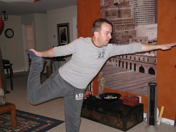
There is often debate about the choice between open and closed-chain exercises for treating knee disorders. One study looked at the benefits of each method for chondromalacia; however, the authors identified chondromalacia as "anterior knee pain," so the participants did not truly have chondromalacia (big surprise). The CKC group performed partial squats, whle the OCK group performed SLR. Each group performed their exercises 20x, twice every day for 3 weeks. Every 2 days, an additional 5 reps would be performed. The results found both groups improved upon pain, but the CKC group had significantly greater improvements in thigh circumference, Q-angle, crepitus, and muscle force (Bakhtiary & Fatemi, 2008). Some potential adjuncts to exercise may include utilization of NSAIDs, injections, and warming needling. If the chondromalacia is not complex, NSAID's have been shown to be beneficial. The more degradation that has occurred in the cartilage, the more invasive treatment becomes. Typically patients with more complex chondromalacia are given a viscosupplementation injection to help increase viscosity and lubrication within the knee joint. Dry needling is becoming a more prominent treatment option for many pain disorders. The theory is that with application of needles, endorphins in the CSF and serotonin in the peripheral blood have increased levels, leading to an analgesic effect. Ling et. al found that warming needling combined with rehabilitation training was more effective in reducing long-term pain than rehabilitation training plus NSAIDs. Surgical Management: When conservative measures fail, surgery is typically the next course of action. The surgical procedure is dependent upon the stage of chondral disease with which the client presents. If the patient has patellar tilt (Outerbridge Level I or II*) The goal of this surgical operation is to restore proper tracking of the patella. Another common surgical procedure is a tibial tubercle osteotomy. The osteotomy attempts to normalize the tibial tubercle to trochlear groove distance. By achieving this, normal congruency is achieved at the patellofemoral joint and less contact stress is induced. One study looked at the effects of arthroscopic debridement for chondromalacia grades III & IV (Outerbridge levels). The treatment involved shaving the remaining cartilage until bleeding bone was reached. Additionally, all the joints were lavaged, and intra-articular debris was removed; this included partial meniscectomies. Following surgery, patients would assume partial weight-bearing on crutches for 5 days before being progressed to full weightbearing. The authors were unable to provide any true indications for when arthroscopic debridement should be performed in grade III & IV chondromalacia, but they still believe it is indicated for certain populations to prolong additional surgery (van den Bekerom et. al, 2007). Given the lack of findings, the evidence is questionable. Other surgery techniques you may see include Bone Marrow Stimulation (Microfracture begins the healing process by making perforations in the subchondral bone), Autologous Chondrocyte Implantation, Osteochondral Grafting, and Patellofemoral Prosthetic Arthroplasty (for patients too young for a TKA). Macmull et. al looked at autologous chondrocyte implantation versus matrix-assisted chondrocyte implantation for chondromalacia. The study found that both methods improved patient symptoms and function, but matrix-assisted chondrocyte implantation was more effective (Macmull et. al, 2012). Again, the type of surgery is largely based off the degree of articular cartilage degradation and other biomechanical influences. Regardless of the surgical technique, patients will go through a strict physical rehabilitation process afterward. A potentially overused treatment for chondromalacia (and patellofemoral pain syndrome) is surgical release of the lateral retinacular fibers. In one study, patients with Q angle >20 degrees did not benefit by lateral release as much as patients with normal Q-angle. The theory is that lateral retinacular fiber release can decrease some of the lateral tracking forces of the patella. Additionally, severing the nerve endings in the internal retinaculum may explain some of the analgesic effects (Vaatainen et. al, 1994). Another study found lateral retinacular release to be as effective as lavage and may be appropriate for patients with patella decentration 5-10 mm and appropriate pain symptoms (Kruger et. al, 2002). Given the conflicting findings, lateral retinacular release should not be an initial course of action. *Outerbridge Classification is used to grade amount of articular cartilage arthritis. (Chevestick 2012) Grade 0: Normal Cartilage Grade 1: Cartilage with softening and swelling Grade 2: Partial Thickness defect with fissures on the surface that do not reach subchondral bone or exceed 1.5 cm in diameter Grade 3: Fissuring to the level of subchondral bone in an area with an area >1.5 cm in diameter Grade 4: Exposed subchondral bone. 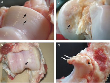
References:
Bakhtiary AH, Fatemi E. "Open versus closed kinetic chain exercises for patellar chondromalacia." Br J Sports Med. 2008 Feb;42(2):99-102. Web. 9 Nov 2012. Brotzman, S., & Wilk, K. Clinical Orthopaedic Rehabilitation. 2nd ed. Philadelphia: Mosby, 2003. 320. Print. Chevestick, A, et al. "Anterior knee pain: the pitfalls of plica and chondromalacia patellae." Advance Newsmagazines. 2.6 (2012). Web. 7 Nov. 2012. Dehaven K, Dolan W, & Mayer P. "Chondromalacia patellae in athletes: Clinical presentation and conservative management." Am J Sports Med. 1979 Jan-Feb;7(1):5-11. Web. 8 Nov 2012. Dehaven KE, Dolan WA, Mayer PJ. "Chondromalacia patellae and the painful knee." Am Fam Physician. 1980 Jan;21(1):117-24. Web. 9 Nov 2012. Doberstein ST, Romeyn RL, Reineke DM. "The diagnostic value of the Clarke sign in assessing chondromalacia patella." J Athl Train. 2008 Apr-Jun;43(2):190-6. Web. 9 Nov 2012. Gomoll, Adreas, et al. "Treatment of Chondral Defects in the patellofemoral joint." Journal of Knee Surgery. 19.4 (2006). Web. 7 Nov. 2012. Harman, Mustafa, et al. "MR Arthrography in chondromalacia patellae diagnosis on a low field open magnet system." Journal of Clinical Imaging. 27 (2003). Web. 8 Nov. 2012. Krüger T, Göbel F, Huschenbett A, Hein W. "Significance of lateral release in the therapy of patellar chondromalacia." Zentralbl Chir. 2002 Oct;127(10):900-4. Web. 9 Nov 2012. Ling Q, Min Z, Ji Z, Le-nv G, Da-wei C, Jun L, Jia-yi S, Ling W, Jin-yan Y, Le-ping H, & Yang B. "Chondromalacia Patellae Treated by Warming Needle and Rehabilitation Training." Journal of Traditional Chinese Medicine. June 2009; 29: 90-94. Web. 8 Nov. 2012. Macmull S, Jaiswal PK, Bentley G, Skinner JA, Carrington RW, Briggs TW. "The role of autologous chondrocyte implantation in the treatment of symptomatic chondromalacia patellae." Int Orthop. 2012 Jul;36(7):1371-7. Web. 9 Nov 2012. McMullen W, Roncarati A, Koval P. "Static and Isokinetic Treatments of Chondromalacia Patella: A Comparative Investigation." JOSPT. Dec 1990. 12:6: 256-265. Web. 8 Nov 2012. Murphy E, FitzGerald O, Saxne T, Bresnihan B. "Increased serum cartilage oligomeric matrix protein levels and decreased patellar bone mineral density in patients with chondromalacia patellae." Ann Rheum Dis. 2002 Nov;61(11):981-5. Web. 9 Nov 2012. Pihlajamäki HK, Kuikka PI, Leppänen VV, Kiuru MJ, Mattila VM. "Reliability of clinical findings and magnetic resonance imaging for the diagnosis of chondromalacia patellae." J Bone Joint Surg Am. 2010 Apr;92(4):927-34. Web. 9 Nov 2012. Salehi I, Khazaeli S, Hatami P, Malekpour M. "Bone density in patients with chondromalacia patella." Rheumatol Int. 2010 Jun;30(8):1137-8. Web. 9 Nov 2012. Vaatainen U, Kiviranta I, Jaroma H, Airaksinen O. "Lateral Release in Chondromalacia Patellae Using Clinical, Radiologic, Electromygraphic, and Muscle Force Testing Evaluation." Arch Phys Med Rehabil. 1994 Oct;75(10):1127-31. Web. 8 Nov. 2012. van den Bekerom M, Patt T, Rutten S, Raven E, van de Vis H, & Albers G. "Arthroscopic Debridement for Grade III and IV Chondromalacia of the Knee in Patients Older Than 60 Years." J Knee Surg. 2007; 20: 271-276. Web. 8 Nov. 2012. Ye QB, Wu ZH, Wang YP, Lin J, Qiu GX. "Preliminary investigation on the pathogeny, diagnosis and treatment of chondromalacia patella." Zhongguo Yi Xue Ke Xue Yuan Xue Bao. 2001 Apr;23(2):181-3. Web. 9 Nov 2012. Yildiz Y, Aydin T, Sekir U, Cetin C, Ors F, Alp Kalyon T. "Relation between isokinetic muscle strength and functional capacity in recreational athletes with chondromalacia patellae." Br J Sports Med. 2003 Dec;37(6):475-9. Web. 9 Nov 2012. |

Dr. Brian Schwabe's NEW Book in partner with PaleoHacks!

Learn residency-level content on our
Insider Access pages 

We value quality PT education & CEU's. Click the MedBridge logo below for TSPT savings!Archives
July 2019
Categories
All
|
 RSS Feed